Home

tornado rimescolare tromba amygdala coronal section Ispirazione presto Spazzare

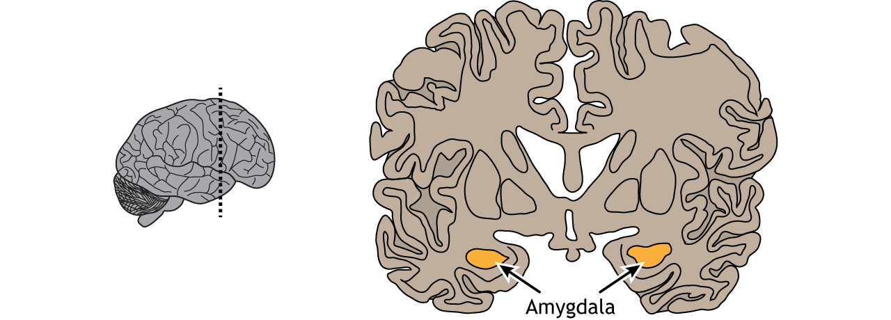
Location of the amygdala in the coronal section of the brain | Medical  student study, Learning science, Brain anatomy
Location of the amygdala in the coronal section of the brain | Medical student study, Learning science, Brain anatomy

Prolonged Attenuation of Amygdala-Kindled Seizure Measures in Rats by  Convection-Enhanced Delivery of the N-Type Calcium Channel Antagonists  ω-Conotoxin GVIA and ω-Conotoxin MVIIA | Journal of Pharmacology and  Experimental Therapeutics
Prolonged Attenuation of Amygdala-Kindled Seizure Measures in Rats by Convection-Enhanced Delivery of the N-Type Calcium Channel Antagonists ω-Conotoxin GVIA and ω-Conotoxin MVIIA | Journal of Pharmacology and Experimental Therapeutics

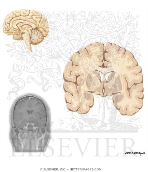
Coronal Section of the Brain (Cut at Basal Ganglia)

Intrinsic Amygdala–Cortical Functional Connectivity Predicts Social Network  Size in Humans | Journal of Neuroscience
Intrinsic Amygdala–Cortical Functional Connectivity Predicts Social Network Size in Humans | Journal of Neuroscience

Thalamic and amygdala–hippocampal volume reductions in first-degree  relatives of patients with schizophrenia: an MRI-based morphometric  analysis - Biological Psychiatry
Thalamic and amygdala–hippocampal volume reductions in first-degree relatives of patients with schizophrenia: an MRI-based morphometric analysis - Biological Psychiatry

Coronal Section Through The Optic Chiasm Figures 14 and - Cerebral Artery

Coronal section through the anterior commissure, amygdala, septal nuclei,  and optic chiasm. Diagram | Quizlet
Coronal section through the anterior commissure, amygdala, septal nuclei, and optic chiasm. Diagram | Quizlet

Internal brain (Coronal cut) Diagram | Quizlet

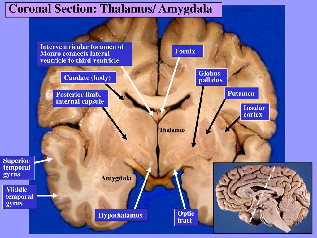
Sectional Anatomy of the Brain - ppt download

Anatomy of the Brain: Coronal Section - TrialExhibits Inc.

Coronal brain sections from control rats without lesions in the... |  Download Scientific Diagram
Coronal brain sections from control rats without lesions in the... | Download Scientific Diagram

Coronal brain sections from rats without lesions (IC, intact control... |  Download Scientific Diagram
Coronal brain sections from rats without lesions (IC, intact control... | Download Scientific Diagram

Differential Effects of Amygdala, Orbital Prefrontal Cortex, and Prelimbic  Cortex Lesions on Goal-Directed Behavior in Rhesus Macaques | Journal of  Neuroscience
Differential Effects of Amygdala, Orbital Prefrontal Cortex, and Prelimbic Cortex Lesions on Goal-Directed Behavior in Rhesus Macaques | Journal of Neuroscience

Coronal Sections Through the Forebrain: Amygdala, Anterior Limb of Internal  Capsule
Coronal Sections Through the Forebrain: Amygdala, Anterior Limb of Internal Capsule

Hippocampus: Anatomy, functions and connections | Kenhub

Figure 8 | Anatomy of the Temporal Lobe

Neural Control of Stress – Foundations of Neuroscience

Neuroanatomical Correlates of Declarative Memory - Human Brain

Duke Neurosciences - Lab 5: Forebrain Sectional Anatomy

Coronal Section of the Brain (Cut at Basal Ganglia) - Dynamic Shapes

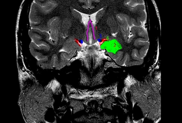
Amygdala anatomy (coronal T2WI MRI) | Radiology Case | Radiopaedia.org

Segmentation: Amygdala

Figure 3 from Neuroanatomy of autism | Semantic Scholar